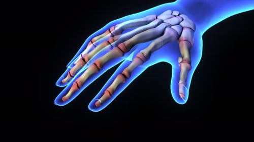
3D Anatomical Animation of Inflamed Finger Joint
By Digital-Display-CompanyOne great value subscription from €14.50/month
- Unlimited downloads of 27+ million creative assets
- AI Tools: video, image and more
- Lifetime commercial license
- Easy cancellation
Already have an account? Sign in
Attributes
- Length
- 0:12
- Resolution
- 3840 x 2160
- File Size
- 15.5 MB
- Frame Rate
- 29.97 fps
- Alpha Channel
- No
- Looped
- No
- Video Encoding
- H.264
- Orientation
- Horizontal
- Commercial License
- Further Information
Description
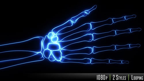
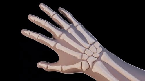
The camera executes a smooth, detailed zoom into a semi-transparent human hand, progressively revealing the intricate skeletal structure beneath the surface. As the perspective shifts, the focus narrows onto a specific finger joint, where vivid red, textured overlays represent acute inflammation or tissue damage. This anatomical visualization utilizes a sophisticated 3D rendering style, characterized by realistic lighting on the bone surfaces and a deep blue, textured background that ensures the primary subject remains the center of attention. The animation is steady and deliberate, effectively guiding the viewer's eye to the site of the pathology. This visual tool is exceptionally well-suited for medical professionals, educators, and content creators specializing in healthcare and biology. It offers an effective way to illustrate complex conditions such as rheumatoid arthritis, joint trauma, or chronic inflammation within a clinical or educational setting. By providing a clear, high-quality representation of musculoskeletal issues, the clip helps to simplify difficult medical concepts for patient education, instructional documentaries, or pharmaceutical marketing materials that require high-end scientific accuracy and professional presentation.