3D Medical Animation of Virus Attacking Cell and Antibody Interaction
By Digital-Display-CompanyOne great value subscription from €14.50/month
- Unlimited downloads of 27+ million creative assets
- AI Tools: video, image and more
- Lifetime commercial license
- Easy cancellation
Already have an account? Sign in
Attributes
- Length
- 0:36
- Resolution
- 3840 x 2160
- File Size
- 45 MB
- Frame Rate
- 30 fps
- Alpha Channel
- No
- Looped
- No
- Video Encoding
- H.264
- Orientation
- Horizontal
- Commercial License
- Further Information
Description
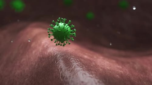
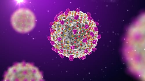
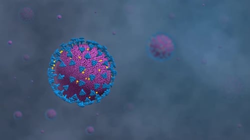
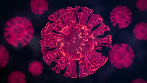
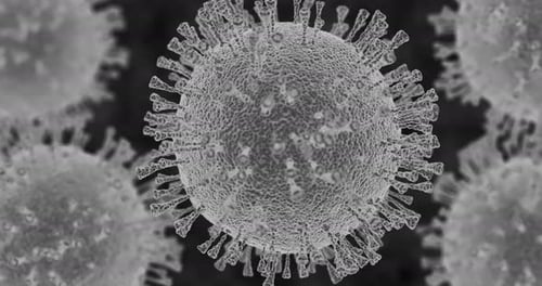
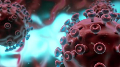
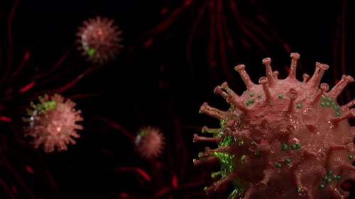
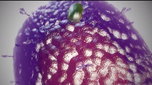
This detailed 3D illustration provides a visual representation of a viral infection process at a microscopic level. The sequence begins with a single virus particle—rendered in deep purples and blues with prominent surface spikes—rotating in a neutral space. The animation transitions to show the virus interacting with a textured, organic environment representing a host cell membrane. As the virus approaches the tissue, glowing green Y-shaped antibodies begin to bind to the viral surface proteins, demonstrating a biological immune response. The camera follows the virus as it docks with specific yellow receptor sites on the cell surface, leading to a dynamic sequence depicting viral entry and fusion. The final scenes show multiple viral particles moving across the host cell surface, illustrating the replication cycle. With smooth camera movements and a professional aesthetic, this clip serves as a visual aid for presentations, educational content, or scientific media focused on virology, immunology, and healthcare. The lighting and textures emphasize the biological structures, making it a clear tool for explaining complex medical concepts.